Part 1: Imaging
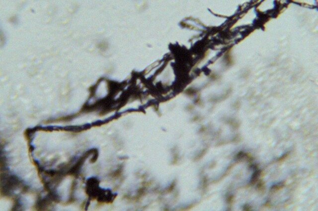
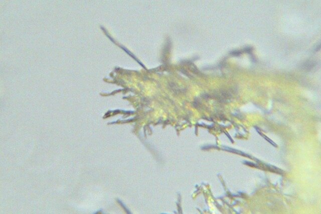
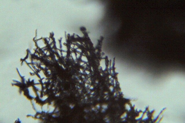
For part 1, I chose to examine mycelium. I had been experimenting with growing mycelium for my biomaterial exploration research, and wanted to see the structures on a microscopic scale, using a scientific microscope with 40x-100x magnification.
I collected small samples using tweezers, which was quite challenging given how delicate mycelium is. I assume the sample was still corrupted during this process. A better way to observe mycelium under the microscope would be to grow it directly on slides to preserve the delicate structure of the root system.
I succeeded to observe chunks of the root structure and also small pieces of the substrate on which the mycelium had grown.
Part 2: Design of smFISH / Spatial Sequencing Assay (Computational)
Part 3: smFISH Image Analysis (Computational)