Aromata-supporting material

Group 14.png
 

Freeze-Dried Cell-Free Biosynthesis

Cell-Free(CF) Bio-production

 
18-9-Melinek-opener.jpeg

Fig. 1. Components of a cell-free protein synthesis reaction: (extract, supplements, and a DNA template) with the key reactions that occur when they are combined.

Melinek et al., “Toward a Roadmap for Cell-Free Synthesis in Bioprocessing.”


Freeze-Dried Cell-Free(FD-CR) reactions

 
F1.large.jpg

Fig. 2. BioBitsTM kits: Freeze-dried educational kits. (A) FD-CF demonstrations require only the addition of water to the supplied reactions and incubation for 1 to 20 hours at 25° to 37°C for observation and analysis by students. In contrast, traditional biology experiments require substantial time, resources, and specialized equipment. (B) With the DNA template and any substrate molecules provided with the FD-CF reaction, the students just have to add water to run a number of bioscience activities and demonstrations.

Huang et al., “BioBitsTM Explorer.”


FD-CF Expression of Aroma Compounds

 
Fragrance-generating-enzymes-as-olfactory-outputs-A-Using-FD-CF-reactions-we.png

Fig. 3. Fragrance-generating enzymes as olfactory outputs. (A) Using FD-CF reactions, we manufactured enzymes that can generate various smells from the Saccharomyces cerevisiae acetyltransferase ATF1. (B) Production of fragrance molecules after substrate addition to overnight FD-CF reactions of ATF1, as detected by headspace GC-MS. Values represent averages, and error bars represent SDs of n = 3 biological replicates.

Huang et al., “BioBitsTM Explorer.”


Microfluidic Bioreactor

Cell-Free Microfluidic Bioreactor

 
full-1846.2017.00014-f4.jpg

Figure. 4.Serpentine channel microfluidic bioreactor design for cell-free production of biotherapeutics.

Abeille et al., “Continuous Microcarrier-Based Cell Culture in a Benchtop Microfluidic Bioreactor.”


Microfluidic Bioreactor

 
Bioreactor Ikram Khan.jpeg

Fig. 5.A 3D-printed microfluidic bioreactor for organ-on-chip cell culture.

Image credit: Ikram Khan


Wearable Olfactory CHI

miniaturized aroma release unit

Fig. 6. Modular scent delivery holders, A) one-piece structure, and B) multi-part decorative structure. C) Design explorations of the scent release mechanism based on 1) Angle between the piezo and the tube. 2) Length and shape of the tube, 3) Assembly of multiple scent release, 4) Clip-on accessories or embeddings in jewelry and piercings.

Wang, Amores, and Maes, “On-Face Olfactory Interfaces.”


Olfactory Interface Placements


Fig. 7. Prototypes that we used for the user study. 1) "Glasses" prototype, 2) "Nose" prototype, and 3) Olfactory necklace. Participants wear the PCB board and battery on their left ear for both on-face designs while hooking the holder at the back part of the cloth for the necklace.

Wang, Amores, and Maes, “On-Face Olfactory Interfaces.”


User Experience Results

 
Screen Shot 2021-08-02 at 5.33.00 PM.png

Fig. 8. Likert Scale for 1 = Extremely Inappropriate or Extremely Uncomfortable and 9 = Extremely Appropriate or Extremely Comfort- able. Error bars correspond to ±1 S.D.

Wang, Amores, and Maes, “On-Face Olfactory Interfaces.”

 
Screen Shot 2021-07-31 at 3.45.25 PM.png

Fig. 9. "Moist" - humidity felt on the face, "Smell" - intensity of the smell, "Burst" - visual spray, "Sound" - emitted when a burst is released. Error bars correspond to ±1 S.D. The wearers smelled the fragrance significantly more than the observers for all the prototypes

Wang, Amores, and Maes, “On-Face Olfactory Interfaces.”


Ethics & policy: Interplanetary synbio

christopher-goodman-the-titan-rick-front-design-logo.jpg

Synthetic biology in space

Promising applications of synthetic biology for long duration space travel include a variety of biologically engineered products and biologically aided processes and technologies. While these applications would aid human space exploration, they could result in several unintended consequences in the long run. Therefore, it’s critical that we discuss and set up protocol frameworks in the context of ethical, legal and social realms.

Ongoing R&D for near future applications of synthetic biology in space points to genetically modified plants and microorganisms designed to produce food and medicine. Other applications include bio-mining, carbon capture and air purification, personalized diagnostic tools, biomaterial etc. However, in the future, these application could expand to other GM organisms, including humans, that are more suited to endure multiple stressors. Long term effects of GM organisms in the context of multiplanetary expansion of terrestrial life and interplanetary interactions should therefore be considered.


Governance and policy

In contrast to earth-based synbio, space applications have additional challenges such as those raised by space microbiology and environmental factors, legal complications, planetary protection, lack of decision-making infrastructure(s), long duration human missions, terraforming and the possible discovery of extraterrestrial (ET) life. Until recently, the main actors of space exploration included politicians, scientists, and engineers. However, with privatized companies, such as SpaceX, entering this industry and turning space exploration into a for-profit economy, it’s important that we set up transparent and more robust governance/policy systems. The desired ecosystem should include more diverse disciplines, such as evolutionary biology, ecology research as well as social sciences to define “long-term” goals of the space programs and ensure an ethical future of synbio in space.“This perspective requires subscribing to a new paradigm that no longer sees ‘long-term’ as months or years but rather as time in an evolutionary context”.

Long-term goals could include, but are not limited to:

  • Non-malfeasance

  • Access

  • Planetary environmental protection

potential governance "actions", each presented in four sections. (TBD)

  • Goal: What is done now and what changes are you proposing?

  • Design: What is needed to make it “work”? (including the actor(s) involved - who must opt in, fund, approve, or implement, etc)

  • Assumptions: What could you have wrong (incorrect assumptions, uncertainties)?

  • Risks of Failure & “Success”: How might this fail, including any unintended consequences of “success” of your proposed actions?


References

Race, M., Moses, J., McKay, C., & Venkateswaran, K. (2012). Synthetic biology in space: Considering the broad societal and ethical implications. International Journal of Astrobiology, 11(2), 133-139. doi:10.1017/S1473550412000018

Criscuolo F, Sueur C, Bergouignan A. Human Adaptation to Deep Space Environment: An Evolutionary Perspective of the Foreseen Interplanetary Exploration. Front Public Health. 2020;8:119. Published 2020 Apr 24. doi:10.3389/fpubh.2020.00119

Design: Radiation resistance

1.)  Overview and rationale:

Humans and most organisms, except for some microbial extremophiles, aren’t fit to survive the host of stressors in deep space or on other planets. To survive long duration space exploration or to inhabit planets other than Earth, we need to make organisms that are more suited to endure multiple stressors. Genetic engineering could help us to build the ultimate astronaut. One that is resistant to high radiation(CTNNB1), and bone loss(SOST) due to altered gravity; but also more mentally resilient with genes that make them less prone to anxiety, and cognitively more agile(PDE4B, FOXP2, CCR5, GRIN2B). We could even augment their physical capabilities by adding a gene that gives them six-fingered hands(HOXA11).

High cosmic radiation is one of the most detrimental stressor in deep space, as well as on a planet like Mars that lacks a magnetic field or a thick atmosphere to shield humans and most organisms that have evolved on earth from radiation. Long duration space exploration will require radiation protection measures. Designing radiation resistant organisms could be a method to ensure long term radiation protection in space.

CTNNBI is a gene with radiation resistance properties. This mutation could be applied to humans to make them more fit to survive and thrive in space.

2.)  Genomic sequence: 


3.)  Genome editor design:

CRISPR/Cas9-based

delivery: TBD

screening: TBD


Protein Design: Structural Colors

gif-octopus-blue-ringed.gif

I’ve always been intrigued by vibrant iridescent colors. In nature, iridescence is caused by structural coloration, which is the production of color by nanostructure surfaces that interfere with visible light, sometimes in combination with pigments.

unsplash-image-5VC4thmwMms.jpg
unsplash-image-3awIIec4R1o.jpg
unsplash-image-hjmED1qivmc.jpg

Another fascinating phenomenon is dynamic coloring in some animals including cephalopods such as squid are able to vary their colors rapidly for both camouflage and signaling. The mechanisms include reversible proteins which can be switched between two configurations. The configuration of reflectin proteins in chromatophore cells in the skin of the Doryteuthis pealeii squid is controlled by electric charge. When charge is absent, the proteins stack together tightly, forming a thin, more reflective layer; when charge is present, the molecules stack more loosely, forming a thicker layer. Since chromatophores contain multiple reflectin layers, the switch changes the layer spacing and hence the color of light that is reflected. Additionally, within the chromatocytes, where the pigment resides in nanostructured granules, we find the lens protein Ω- crystallin interfacing tightly with pigment molecules.

Protein Analysis

Pick any protein (from any organism) of your interest that has a 3D structure

Briefly describe the protein you selected and why you selected it.

  • Identity the amino acid sequence of your protein.

    • How long is it? What is the most frequent amino acid?

    • How many protein sequence homologs are there for your protein?

      Hint: Use the pBLAST tool to search for homologs and ClustalOmega to align and visualize them.

    • Does your protein belong to any protein family?

  • Identify the structure page of your protein in RCSB

    • When was the structure solved? Is it a good quality structure?

    • Are there any other molecules in the solved structure apart from protein?

    • Does your protein belong to any structure classification family?

  • Open the structure of your protein in any 3D molecule visualization software

    • Visualize the protein as "cartoon", "ribbon" and "ball and stick".

    • Color the protein by secondary structure. Does it have more helices or sheets?

    • Color the protein by residue type. What can you tell about the distribution of hydrophobic vs hydrophilic residues?

    • Visualize the surface of the protein. Does it have any "holes" (aka binding pockets)?


References

Williams, T.L., Senft, S.L., Yeo, J. et al. Dynamic pigmentary and structural coloration within cephalopod chromatophore organs. Nat Commun 10, 1004 (2019). https://doi.org/10.1038/s41467-019-08891-x

Remote Lab Automation

Instructions

  • Only one student at a time can work on the machine, so first, go to the slack channel #opentron-users and post a message that you are starting a session. Do this every time you plan to connect to the machine, even if it's just for taking an image.

  • You will need to connect to the remote machine using Remote Desktop software. This process is only for registered students, although if you do have access to an OpenTrons machine, feel free to follow along.

    • Windows users: in the search bar type Remote Desktop and run it

    • Mac users: download Microsoft Remote Desktop from the app store and run it

    • Linux users: use any RDP client of your choice.

  • Login to: opentrons.media.mit.edu

    • Username and Password will be given to the registered student over Slack

  • Click on the OpenTrons icon in the lower bar. You should see a single Robot in the list. Connect to it by hitting the toggle switch next to it

  • Scroll down over the list of options on the right until you see the Robots Controls > Light toggle. Turn the lights on if they're off. This is also useful to scare labmates.

  • Minimize the OpenTrons app. You should see two windows open:

    • On the right, Chrome browser shows the live OpenTrons cameras. When you turned the lights off/on, you should see the live feed changing. If for some reason you cannot see this window, open Chrome and in the address bar type localhost and hit Enter

    • On the left, a Microsoft Edge window with the Jupyter notebook. This wondow is where you will execute code to run experiments. If for some reason you cannot see this window, open the OpenTrons app, and scroll to Advanced Settings > Jupyter Notebook, click Open. And then go to HTGAA Logo > Warmup Exercise. Clicking it should open the notebook

  • It's time to execute! But first, Make a Copy of the Jupyter notebook by going to File > Make a Copy. A new tab should appear, change the title of the file so it contains your name, and work only on your file.


Next Generation Synthesis

Part A: Primer Design and Fragment Assembly

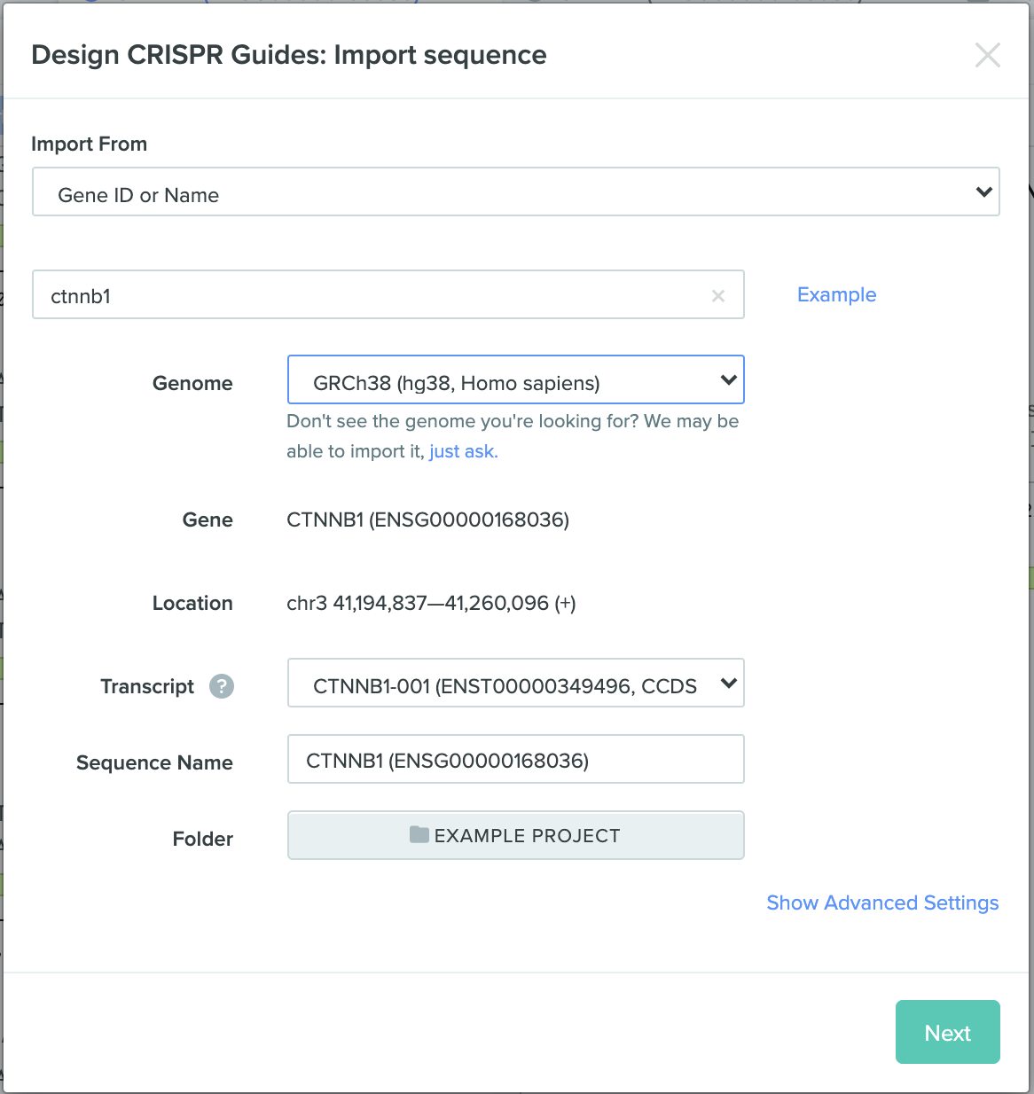
In this part, we will prepare and order the primers that will generate a library of mutated amilCP expressing E. coli cells. We will use Gibson Assembly to insert our mutated gene into a plasmid, which in turn will be transformed into electrocompetent E. coli cells. First, let's understand what is Gibson Assembly and how to design primers for it.

Design primers to amplify two sets of amplicons from mUAV plasmid. The amplicons sets must include one end that overlaps by 20-22 bases with distinct ends of the pUC19 backbone.

  • Restriction digest pUC19 with PvuII and identify the backbone you want to use for your assembly. [Hint: You need a selection marker and origin of replication!]

  • Import the mUAV plasmid sequence into Benchling, by going to Import DNA Sequences > Search External Databases and input the GenBank identifier MG252981.1

  • Interestingly, we will be actually be using a Twist Gene Fragment as the source DNA. For our 1kb fragment, both the price and delivery times are better compared to plasmids, and importantly, require less TA lab work (no need to miniprep). To examine which fragment we ordered, use this link: https://benchling.com/s/seq-uivVVxZrv3WxMWNhyTJu

  • Identify the amilCP gene, RBS, promoter, and terminators in Plasmid mUAV.

  • As described in Liljeruhm et al, the amilCP gene contains a chromophore (CP) region that can be mutated to express different colors. The mutation region is: cagTGTCAGtac

    • Identify the CP mutation sequence (TGTCAG) in the gene and annotate it.

    • Use a codon table and convert the following figure to a table of colors of DNA sequences


    We will split create two fragments out of the mUAV plasmid. One fragment will contain the RBS, promoter and first part of amilCP gene right up until the CP mutation region. The second fragment will include the CP mutation all the until the terminators.

  • To generate these two fragments, we will design four primers (two forward, two reverse). Each of the primer sequences should follow the primer design guidelines to increase your chances of success in the experiment. A few guidelines as taken from here

    • Primer Length: It is generally accepted that the optimal length of PCR primers is 18-22 bp. This length is long enough for adequate specificity and short enough for primers to bind easily to the template at the annealing temperature. However, in our case we will design long overhangs to prepare for Gibson Assembly, so the binding region of the primer should be 18-22 bp, followed by a 20-22 bp overhang.

    • Melting Temperature (Tm): the temperature at which one half of the DNA duplex will dissociate to become single stranded and indicates the duplex stability. Primers with melting temperatures in the range of 52-58C generally produce the best results. Primers with melting temperatures above 65C have a tendency for secondary annealing. Importantly, primers in the same set should have a similar Tm (5C) between each other!

    • GC clamp: The presence of G or C bases within the last five bases from the 3' end of primers (GC clamp) helps promote specific binding at the 3' end due to the stronger bonding of G and C bases. More than 3 G's or C's should be avoided in the last 5 bases at the 3' end of the primer.

    • GC content: the number of G's and C's in the primer as a percentage of the total bases should be 40-60%.

    • Primer Secondary Structures (a.k.a primer dimer): Presence of the primer secondary structures produced by intermolecular or intramolecular interactions can lead to poor or no yield of the product. They adversely affect primer template annealing and thus the amplification. They greatly reduce the availability of primers to the reaction.

      • Benchling allows us to check for secondary structure by selecting part of the sequence > Create Primer > Check Secondary Structure

      • Another great online software is NUPack.

      • It can be quite hard to design primers with no secondary structures. A rule of thumb is to keep the Gibbs free energy of each structure at above -10kcal. For your task, just report what are the secondary structures did you get for your primer pairs.

    • In your report, elaborate how you chose your primers and according to these design guidelines. Notice that we can't always make every primer perfect, but the more guidelines you follow the higher your chances of success.

Primers

  1. Outer Forward Primer:

    • mUAV: identify an 18-22bp region just before the promoter/RBS. Note that you can see your GC content and Tm on the bottom. Right-click and Create Primer (Forward) to examine the design parameters. Copy the sequence to a text editor.

    • pUC19: identify the ~20bp region just after the PvuII cut site.

    • Combine these sequences to get your Outer Forward Primer

    • Make sure your 5 to 3 orientation is right, this is very confusing!

  1. Outer Reverse Primer:

    • mUAV: identify an 18-22bp region just after the terminators. Right-click and Create Primer (Reverse) to examine the design parameters. Copy the sequence to a text editor.

    • pUC10: identify the ~20bp region just after the PvuII cut site.

    • Combine these sequences to get your Outer Reverse Primer

    • Make sure your 5 to 3 orientation is right, this is very confusing!

  1. Inner Reverse Primer:

    • mUAV: identify the chromophore (CP) mutation region

    • Select a 18-24bp region just before the CP mutation region. Right-click and Create Primer (Reverse).

    • Copy the sequence to a text editor to get your Inner Reverse Primer

  1. Inner Forward Primer (Mutations):

    • mUAV: identify the chromophore (CP) mutation region

    • Select a 18-24bp region before and after the CP mutation region, as well the the CP mutation region (total length: 18-24 + 6 + 18-24 = 42-54bp!)

    • This will be our PCR primer + overhang for Gibson Assembly.

    • Copy this sequence to a text editor. Now, use the table you made above to choose which color variant you wish to express.

      • You can have multiple colors together! each E.coli cell will only get one plasmid and express it, but we will have many cells and a variety of colors. You can also order different mixes. Go wild.

      • To make a mutation library, you have to options:

        • Prepare a bunch of primers, that will be synthesized seperately and you will later mix them together using the robot.

        • Use degenerate bases, where a single letter (e.g. H) means it could be a number of different bases (e.g. A/C/T).

        • For example, if we synthesize the sequence HTGAA, we will get a mixture of: ATGAA, CTGAA, TGTAA in a single tube.

    • Your set of sequences is your Inner Forward Primer

This week's task is purely in-silico design. Next week we will use the same primers you designed and perform fragment assembly using the remote robot. We will send your designed primers for DNA synthesis. Please send all of your primers (simply as text files) to Eyal by 03/22 09:00, so we can order them and have the experimental setup ready by next Wednesday. Your number of primers can be between four to ten per student. Notice that if you use degenrate bases, you can have more than "one" primer sequence that still counts as a single order. For example, ordering HTGAA will give you a single tube that contains three primers: ATGAA, CTGAA, TGTAA

Final Projects and Twist Genes

In this exercise, we are extracting a specific gene (amilCP) from a plasmid and mutating it using PCR. As we talked about in class, Next Generation DNA synthesis is changing the way we think about bio-design. Twist Bioscience, as part of its gracious support to HTGAA, offers us a special budget for ordering gene fragments and clonal genes. These could be extremely handy for your final projects. Essentially, you could choose any gene you fancy and submit it to Twist. When ordering clonal genes, they already take care of the work of inserting it into a vector (plasmid). Meaning, you can just choose a gene and choose an bacterial expression vector and you will get a bacteria expressing your synthetic gene (with no lab work!)

Measurement and Imaging: Mycelium

Part 1: Imaging

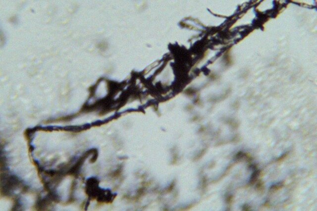
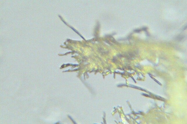
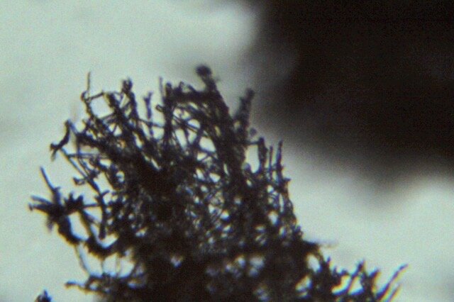
For part 1, I chose to examine mycelium. I had been experimenting with growing mycelium for my biomaterial exploration research, and wanted to see the structures on a microscopic scale, using a scientific microscope with 40x-100x magnification.

I collected small samples using tweezers, which was quite challenging given how delicate mycelium is. I assume the sample was still corrupted during this process. A better way to observe mycelium under the microscope would be to grow it directly on slides to preserve the delicate structure of the root system.

I succeeded to observe chunks of the root structure and also small pieces of the substrate on which the mycelium had grown.

Part 2: Design of smFISH / Spatial Sequencing Assay (Computational)

Part 3: smFISH Image Analysis (Computational)

Circuits, Sensors, & Cell-Free Systems

Part I - in silico homework

You will design a useful synthetic minimal cell.

1. Pick a function.
2. Design all components that would need to be part of your synthetic cell.
3. Experimental details

An example solution given below, based on: Lentini, R. et al., 2014. Nat comm, 5, p.4012.

1. Pick a function.

1A What would your synthetic cell do? What is the input and what is the output.
Expand the sensing capacity of bacteria. Input: theophylline (inert to bacteria). Output of the SMC: IPTG. Output of the whole system: GFP produced in bacteria.

Theophyline Aptamer reference: Martini, L. & Mansy, S.S., 2011. Cell-like systems with riboswitch controlled gene expression. Chemical Communications, 47(38), p.10734.

1B Could this function be realized by cell free Tx/Tl alone, without encapsulation?

No. If the IPTG was not encapsulated, it would go into the bacteria without the need of theophylline-induced membrane channel synthesis, thus the synthetic cell actuator would not exist.

1C Could this function be realized by genetically modified natural cell?

Yes, in this particular case: the theophylline aptamer could be incorporated into transformed gene. This lacks the generality though – it is easier to make SMC than modify bacteria, so in this system single bacteria reporter can be used to detect various small molecules.

1D Describe the desired outcome of your synthetic cell operation.
In presence of SMC, bacteria sense theophylline.

2. Design all components that would need to be part of your synthetic cell.

2A What would be the membrane made of?
Phospholipids + cholesterol.

2B What would you encapsulate inside? Enzymes, small molecules.
Cell free Tx/Tl system, IPTG, gene for membrane transporter under the control of theophylline aptamer.

2C Which organism your tx/tl system will come from? is bacterial OK, or do you need mammalian system for some reason? (hint: for example, if you want to use small molecule modulated promotors, like Tet-ON, you need mammalian).

Bacterial, because of the theophylline riboswitch used as SMC input.

2D How will your synthetic cell communicate with the environment? (hints: are substrates permeable? or do you need to express membrane channel?)

The membrane is permeable to the input molecule (theophylline), the output is IPTG that will cross the membrane via the membrane pore created after theophyline-initiated gene expression.

3. Experimental details

3A List all lipids and genes (bonus: find the specific genes; for example, instead of just saying “small molecule membrane channel” pick actual gene)

Lipids: POPC, cholesterol

Enzymes: bacterial cell free tx/tl

Genes: a-hemolysin (aHL) to encapsulate in SMC,

Biological cells: E.coli transformed with GFP under T7 promoter and a lac operator

3B How will you measure the function of your system?

Measure GFP output of the cells, via flow cytometry. Alternatively, use enzymatic reporter, like luciferase, and measure bulk output of the enzyme.

Part II - Experimental homework

For this week’s experimental homework, you get to test different hypotheses about transcription and translation reactions.

Cell-free transcription

You will design a cell-free transcription reaction where the transcription mix produces fluorescent RNA aptamer called Broccoli, see this work by Filonov et al. The RNA is an aptamer that enhances the fluorescence of a small molecule ligand. Fluorescent RNA aptamers. In the transcription reaction, the Broccoli RNA should fold into a conformation which binds a ligand. The ligand--DFHBI--will fluoresce upon binding to the folded Broccoli. Erkin will set-up your designed experiments for you, following the basic recipe below. He will then measure the aptamer fluorescence increase over time using a 96 well plate to check if the mRNA is successfully expressed in the cell-free system. He will share the data with you and you will interpret the results. 

This is a good place for you to generate a hypothesis and for Erkin to test your hypothesis. You can try up to 7 condition (perhaps more if the reagents allow it) plus a positive control. You are encouraged to sample different conditions involving the following antibiotics:

-Kanamycin
-Ampicillin
-Rifamycin
-Puromycin
-Vancomycin
-
 Dactinomycin
-Chloramphenicol

For example, you might want to sample different antibiotics or different concentrations of select few (0.1X, 1X, 10X, etc)

In addition, you might want to play around with the working stock concentrations of the basic kit. For example, transcription reaction includes Magnesium and Potassium salts. Would too little (or too much) of those hurt your reaction?  You can also test the effects of different pH (by adding a base or an acids into the reaction) or effects of high energy light exposure (i.e, UV light). You might even ask Erkin to spit into a well. Finally, you can also test temperature effects. The key enzyme in this transcription reaction come from a bacteriophage virus which preys on bacteria that like to reside in our bodies. What would the optimal temperature for this reaction be? How would you test that?

Explain in your webpage the importance of each of the reagents you used to transcribe mRNA using the cell-free system. Think about why including controls in your experiments are important and what your positive control in your experimental design would be. Read about antibiotics in general. What makes a molecule an antibiotic? What are the different modes of action of antibiotics? Interpret the results of your experimental results and compare to your initial hypothesis. What would do differently the next time?

Cell-free transcription and translation

First, Erkin will set up a cell-free protein expression experiment using three different cell-free transcription translation systems as shown below. The template (plasmid) is GFP and its spectrum. Once the reaction is setup, Erkin will incubate the reaction at 30C.

1- PURExpress® In Vitro Protein Synthesis Kit from NEB that is prepared roughly according to the protocol based in this seminal Shimizu et al. paper.

2- BioBits®: Central Dogma Kit that is prepared roughly according to the protocol based in this paper by Huang et al. and stored in a -20oC freezer as a lyophilate for 4 weeks.

3- Homemade cell extract that was stored in a -80oC freezer for less than a week. This system also requires the addition of energy and amino acid mixtures (that are already present in the other two systems above). These mixtures were prepared roughly according to the protocol based in this paper by Sun et al, while the extract was made by sonication according to protocol based on the this paper by Kwan et al.

Think and read about the relationship of transcription to translation. Why are these reactions essential to all life called what they are called? Remind yourself the Central Dogma. Explain in your webpage the importance of each of these reagents used to transcribe mRNA and translate proteins using the cell-free system. Come up at least one advantage of cell-free systems over working with live cells. Bet on which of the three systems will work better. Why did you choose one over the other? Think about how ‘working better’ would look like in this experiment. What would be your output from this experiment? How would you quantify it?

Second, you will again have the chance to generate and test different hypotheses here.
For this, we will mainly utilize 
BioBits®: Central Dogma Kits Try to design your experiments such that you will be testing a hypothesis. You can try up to 7 condition plus a positive control. Again, you are encouraged to sample different conditions. These include the list of antibiotics and conditions mentioned above. If you decide to test the effects of different reaction components refer to the table above which Erkin will set up using the homemade cell extract.

Think about what your positive control in your experimental design would be. Come up with at least one good negative control. Interpret the results of your experimental results and compare to your initial hypothesis. What would do differently the next time?